


myocardial perfusion imaging(188)
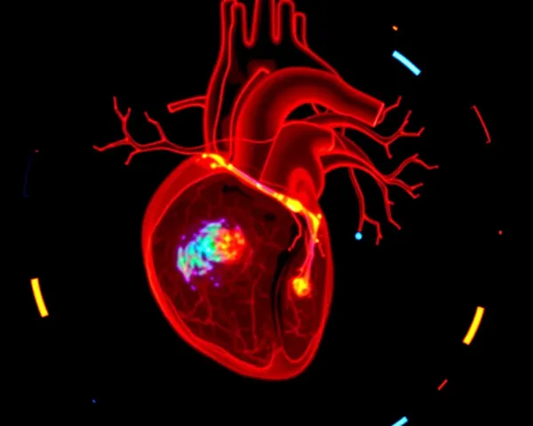
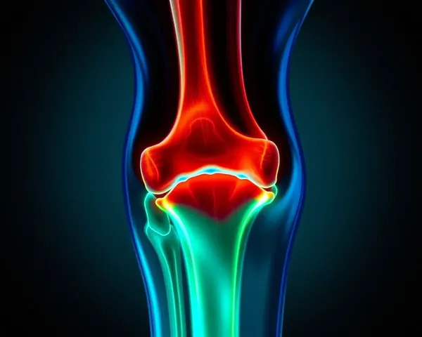
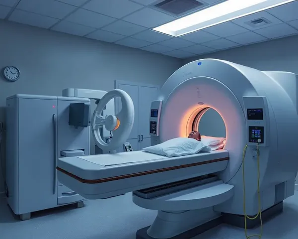
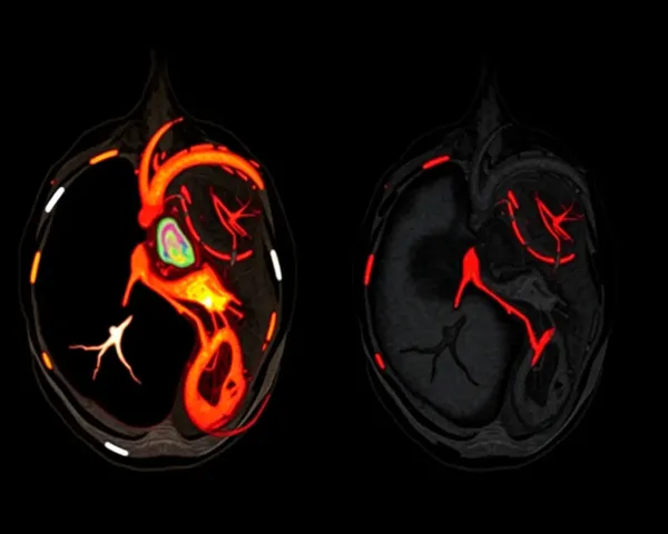
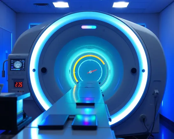
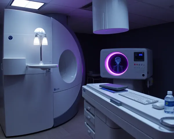
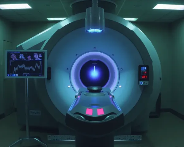
Myocardial perfusion imaging is a non-invasive diagnostic test that uses small amounts of radioactive tracers to visualize the flow of blood to the heart muscle, allowing doctors to assess the health of the heart's blood vessels and identify any areas of reduced blood flow, which can indicate coronary artery disease, heart attack, or other cardiovascular conditions.


- 1
- 2
- 3
- 4
- 7
Top Recommended Prompts
Create a medical illustration of a patient undergoing a stress test, with a technician administering a medication to induce stress, and a computer screen displaying the myocardial perfusion imaging results, highlighting areas of reduced blood flow.
Design an infographic explaining the principles of myocardial perfusion imaging, featuring a diagram of the heart with labeled coronary arteries, a pie chart illustrating the percentage of blood flow to different areas, and a key highlighting the importance of this diagnostic technique.
Generate an illustration of a cardiac MRI scan showing the visualization of myocardial perfusion imaging, highlighting the blood flow and oxygen supply to the heart muscle, with detailed annotations of the coronary arteries and surrounding tissues.

