


magnetic resonance imaging knee(198)
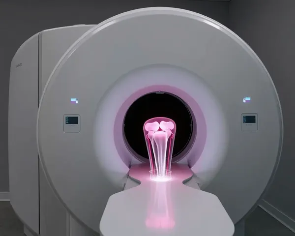
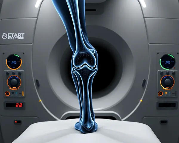
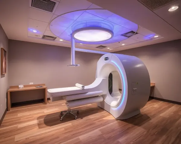
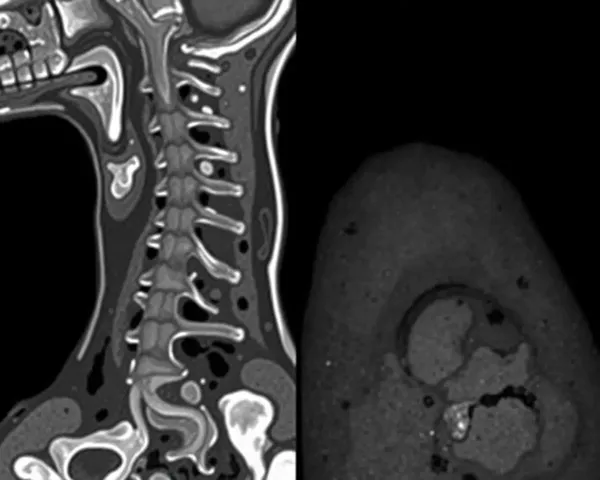
Magnetic Resonance Imaging (MRI) of the knee is a non-invasive medical imaging technique that uses a strong magnetic field and radio waves to produce detailed cross-sectional images of the knee joint, allowing for the diagnosis and monitoring of conditions such as osteoarthritis, ligament tears, and meniscal damage, as well as the detection of bone and soft tissue injuries, and guiding surgical procedures.


- 1
- 2
- 3
- 4
- 7
Top Recommended Prompts
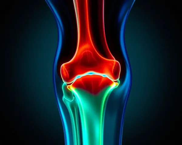
A magnetic resonance imaging (MRI) scan of a knee joint with osteoarthritis, depicting the degenerative changes in the articular cartilage, bone, and surrounding soft tissues, including the menisci and ligaments.
An MRI image of a knee joint with a torn anterior cruciate ligament (ACL), highlighting the damage to the ligament and surrounding soft tissues, with a focus on the ligament's disruption and surrounding inflammation.
A high-resolution magnetic resonance imaging (MRI) scan of a healthy adult human knee joint, showcasing the intricate details of the joint's structure and surrounding tissues, including the patella, quadriceps tendon, and ligaments.

