


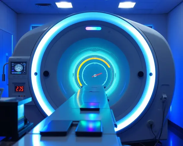
tomographic images(1,454)
Tomographic images are two-dimensional representations of internal structures within a body created by reconstructing data obtained from various imaging modalities, such as X-rays, computed tomography (CT), magnetic resonance imaging (MRI), or positron emission tomography (PET), allowing for non-invasive visualization of internal organs, tissues, and abnormalities.


- 1
- 2
- 3
- 4
- 49
Top Recommended Prompts
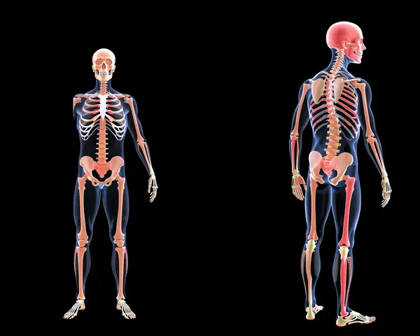
Tomographic images of the skeletal system, showcasing the detailed anatomy of the bones, joints, and surrounding soft tissues, highlighting potential injuries or conditions such as osteoporosis or arthritis.
High-resolution tomographic images of the lung, revealing the intricate branching patterns of the bronchi and alveoli, highlighting the effects of chronic obstructive pulmonary disease (COPD) on lung tissue.
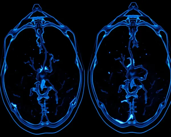
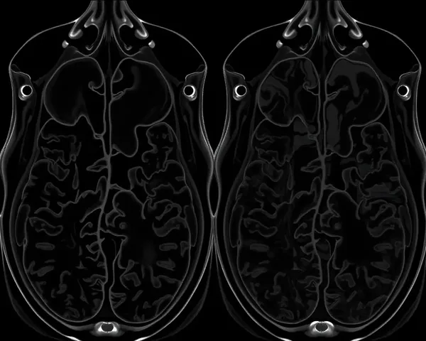
Tomographic images of the human brain showcasing the intricate neural networks and blood vessels, highlighting the complex relationships between different brain regions and their functions.

